IMAGEN MÉDICA ESPECIALIZADA DE PUERTO PEÑASCO
Calidad en imagen al servicio de usted
imagenpenasco@gmail.com
638 105 1906
638 688 3049
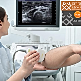
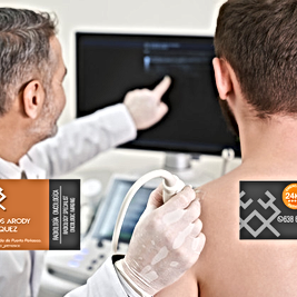
ULTRASONIDOS
ULTRASOUNDS (ENG)
SPORT MEDICINE
PLASMA RICO EN PLAQUETAS
PRP PLASMA RICH PLATETEL
IMAGEN MEDICA
Más
---
----